The idea of shooting yourself with radiation sounds scary, but x-rays are very safe. The American College of Radiology recommends a limit of 100 mSV of exposure to diagnostic radiation — equal to 10,000 chest x-rays.
Want a bit more peace of mind? Hopefully, understanding x-rays can help ease your fears. Read on to learn all about the history of x-rays, what x-rays are, and how an x-ray machine works.
History of the X-Ray
X-rays were accidentally discovered by Wilhelm Roentgen in 1895. The German physicist was experimenting with electron beams inside of a gas discharge tube. When the beam was on, his lab’s fluorescent screen would glow.
Fluorescent things usually glow when exposed to electromagnetic radiation. But he was surprised the heavy cardboard around the tube didn’t block the radiation. This was the first discovery of x-rays.
He kept placing various objects between the tube and the fluorescent screen to see if anything would block the radiation. When he placed his hand in front, his bones were projected onto the screen. So, he didn’t only discover x-rays but also realized their practical medical application.
What is an X-Ray
So, what’s an x-ray? Once again, it’s a form of electromagnetic radiation. It’s not all that different from visible light, except its wavelength is too short for the human eye to detect it.
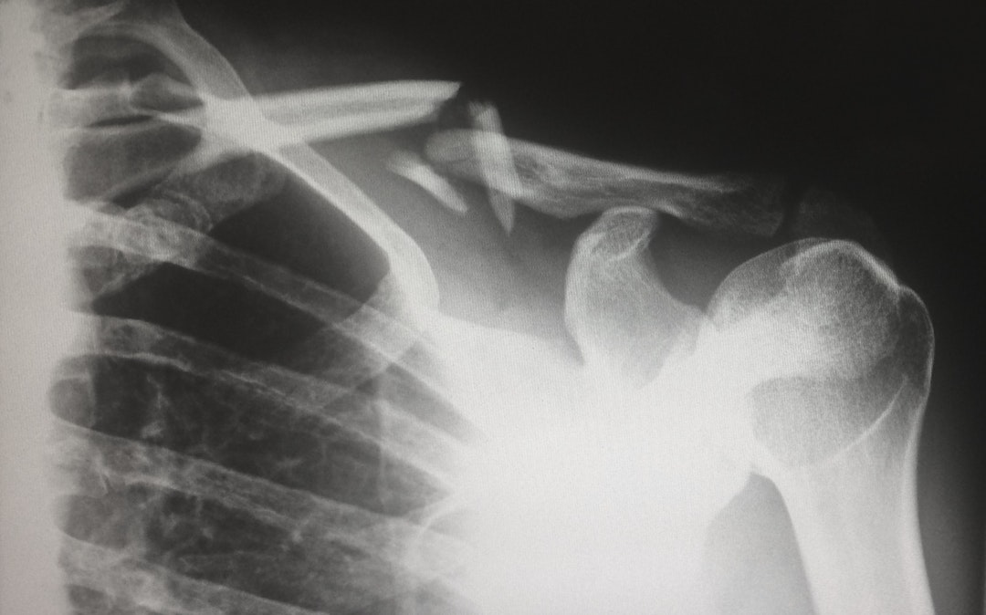
This shorter wavelength means that x-rays have higher energy. So they’re capable of passing through most objects, including human tissue. That’s why we can see the bones within the human body.
If there’s a detector on the other side, all x-rays that pass through the body appear as a black shadow on the x-ray film. Anything that absorbs the x-ray appears white — i.e. your bones.
How an X-Ray Machine Works
The X–ray machine is an invention that includes x-rays in it. It is usually operated by X–ray technicians. X–ray technicians are highly demanded now. Well becoming an x–ray tech can be very useful. For your information, X–Ray techs make around $63k a year. Coming back to the main point – how does an x–ray machine work? Well, at the core of an X–ray machine is an electrode pair inside a glass vacuum tube — very similar to Roentgen’s original experiment.
The negatively charged cathode is a filament heated by the x-ray machine’s current. This causes electrons to come off of the filament’s surface. The positively charged tungsten anode attracts the electrons across this tube.
This process creates enough energy to create an x-ray in the form of a high energy photon. The thick lead contains the photons, but a small window lets some escape out via a narrow beam and onto the patient.
A camera receives this pattern of x-ray light and develops film, like any other camera. But instead of visible light, a chemical reaction takes place. Areas exposed to more x-rays appear darker, which are your air-filled cavities, while hard materials like bone appear white.
Usually, once the process is all said and done, you can access your x-rays if your doctor uses a doctor-patient interface like Medinformatix. How cool is it that you can get a view of the inside of your body?
It is also possible to view some soft tissue using an x-ray. To do this, contrast media is put into the body. These are liquids that absorb x-rays better than soft tissue on its own.
The patient swallows a barium compound so organs, the endocrine system, and the digestive system can absorb x-rays. Or the doctor injects the media into veins so the circulatory system becomes visible. This can be particularly helpful for diagnosing lung cancer and other types of soft-tissue cancers.
Now You Know How X-Rays Work!
We hope this informative guide has taught you all you want to know about x-rays and how an x-ray machine works. Interested in learning about the most cutting-edge medical technology? Read all about how Robotic Process Automation can increase efficiency in healthcare.


